Intervertebral Disc Changes after Vertebral Distraction Performed During Posterolateral Spine Fusion for Lumbar Segmental Instability
Phan Dinh Qui Du,Charlie Arendt, Stig M Jesperesen and Tamás S Illés
DOI10.21767/2471-8173.100015
Phan Dinh Qui Du1, Charlie Arendt1, Stig M Jesperesen2 and Tamás S Illés1,2,3*
1Department of Orthopedics and Traumatology, Brugmann University Hospital, Université Libre de Bruxelles, Belgium
2Department of Orthopedic Surgery and Traumatology, Odense University Hospital and Institute of Clinical Research University of Southern Denmark, Odense, Denmark
3National Medical Academy, Paris, France
- *Corresponding Author:
- Tamás S Illés
Department of Orthopedics and Traumatology, Brugmann University Hospital, Université Libre de Bruxelles, Place Van Gehuchten 4 1020 Brussels, Belgium
Tel: 32 24773080
Fax: 3224772161
E-mail: tamas.illes@chu-brugmann.be
Received date: March 14, 2016; Accepted date: March 28, 2016; Published date: April 02, 2016
Citation: Qui Du PD, Arendt C, Jesperesen SM, et al. Intervertebral Disc Changes after Vertebral Distraction Performed During Posterolateral Spine Fusion for Lumbar Segmental Instability. Spine Res. 2015, 2:1. doi:10.21767/2471-8173.100015
Abstract
Analysis of intervertebral disc regeneration after disc height restoration by posterolateral spinal fusion with lumbar distraction of 68 patients diagnosed with degenerative lumbar instability.
Keywords
Disc degeneration; Instability; Posterior fusion; Disc height restoration; Disc regeneration
Introduction
Degenerative disc disease (DDD) is the main cause of low back pain [1,2]. The most important factor involved in the development of DDD is the vertical load of spine, which can activate enzymatic processes facilitating discs degenerations, in addition to the purely direct (increased fluid stress and hydrostatic pressure) and indirect (decreased permeability and nutrition) mechanical effects. [3] The repetitive flexion movements and certain genetic characteristics may also play an important role in DDD. [4].
One of the most common conditions accompanying DDD is segmental instability of the spine. The pathogenesis of segmental instability was first described by Kirkaldy-Willis [5]. He divided the evolution of the disease into three stages called temporary dysfunction, unstable phase and secondary stabilization.
In temporary dysfunction, early signs of disc degeneration can only be discovered by MRI. The abnormality compared to normal discs is a low signal in the nucleus pulpous without decreased disk height or changes in the contour of the annulus fibrous. These abnormalities are often referred as “dehydrated”, “desiccated” discs, or simply as “dark discs” [6]. In spite of the early radiological signs of disc degeneration, many patients do not show any symptoms thus the clinical significance of dark disks is quite unknown [7].
Disc degeneration process continues in the second, unstable phase, causing the change in the structure of collagen fibers, reducing proteoglycan and, consequentially, water concentration, which unequivocally leads to the loss of disc flexibility. These changes lead to height reduction of discs leading to laxity of restraining structures that result in the instability of the spine segment affected [8].
The clinical symptoms of DDD are diverse, so diagnosis is often inaccurate [9]. The main symptom is the pain, which can arise from many reasons and in many different forms. Pain could mostly arise from endplate degeneration, small joint arthritis, and nerve compression caused by disc protrusion, and inflammation caused by the release of biochemical mediators [10]. Segmental instability of the lumbar spine can be a significant source of pain, as well, although there is not an unequivocally accepted theory concerning the connection between instability and lumbar spine pain. As a result of the progression of instability, movements of the spine become anomalous, exaggerated, even limited [11]. Repetitive hypermobility can cause degenerative spondylolisthesis, and in an extreme case, even degenerative scoliosis [10].
Clinical appearance of the instability is not specific either. Next to spine pain, no clinical signs or symptoms are known which can unequivocally be connected to lumbar spine instability. Despite more and more advanced imaging techniques there was no success in proving a connection between radiological signs and clinical symptoms of the instability [12].
The lumbar instability does not even have a definition collectively accepted by all specialists working with diagnosis and treatment of the disease. On the other hand, most agree on the existence of lumbar instability (developing in DDD) and the pain it causes [8].
Similar to its diagnosis, there is no unequivocally accepted recommendation for the treatment of lumbar instability. Conservative treatment is the traditional approach, which is usually a combination of painkillers and physiotherapy. In the case of ineffectiveness, a surgical procedure is a viable alternative. Most of the surgical procedures aim to use spine fusion to mend the pain caused by disc degeneration after sacrificing the intervertebral discs. Many techniques and procedures were developed to attain spine fusion, in most cases; however, the actual surgical approach is determined by the surgeon’s experience and opinion [13,14].
Promotion of disc regeneration may be a completely new therapeutic approach. Disc cell therapies, in which cells are injected into the degenerate disc to regenerate the matrix and restore function, seems to be an attractive, minimally invasive method of treatment. The repair process in human discs, however, is a very slow process, even in healthy human discs.
Considering that disc reparation that relieves clinical symptoms requires a long time, quality of life is questionable during this lengthy process [15].
Even though animal studies have been demonstrated that disc regeneration can be enhanced by pressure reduction, there has been no surgical instrumentation developed to help reducing disc pressure [16,17].
The aim of the present study was to observe intervertebral disc behavior after performing posterior intervertebral distraction during lumbar spinal fusion carried out in cases of segmental lumbar instability. The influence of posterior distraction on lumbar lordosis and its effect on patient satisfaction after surgery was also studied.
Materials and Methods
68 patients (36 women and 32 men) operated by the same surgeon were selected for this study based on the protocol approved by the ethical committee of the institutions of the senior author. The mean age of patients was 49.2 years (SD: 10.0) at the time of surgery. The minimal follow-up is two years, the maximal is 9.4 years. The average follow-up is 7.5 years (SD: 1.7). Indication for lumbar spine fusion surgery was degenerative segmental instability, with a high percentage of low back pain according to the patient’s Oswestry disability index (ODI) [18].
The instability was diagnosed both functionally - brace wearing and preoperative imaging techniques such as plain radiographs and MRI. The surgery was performed only if the pain has been significantly reduced according to the patients’ considerations after three months of continuous lumbar brace wearing. Patients were excluded if they had signs of neural compression, generalized disc degeneration showed on radiographs or other specific radiological findings such as stenosis, spondylolisthesis (more than Grade I), fracture, infection, or neoplasm.
All patients underwent posterior spinal fusion surgery using Claris transpedicular screw and rod system (ScientX France, than AlphatechSpine, USA). In each case, the transpedicular screw insertions were followed by distraction on the pre-bended titanium rods. The rod bending depended on the degree of lordosis desired to be achieved postoperatively while the extent of distraction was defined by the average height of healthy discs above and below the planned fusion segments. After the correction maneuver large amount of bone allograft transplantation from the local bone bank was performed posterolaterally to achieve good bone fusion.
Study protocol: Measurement values and methods
The same imaging protocol was used pre and postoperatively. Plain radiographs and MRI scans were performed in each case. To evaluate changes in he sagittal balance after the posterolateral fusion, lumbar lordosis was measured both preand postoperatively.
The posterior intervertebral space was measured twice in the area connecting the tip of the posterior margin of the vertebral body on each radiographic image and the mean values were used to determine relative disc space changes in percentages. Changes in the total length of the lumbar spine were also measured, using a line is drawn next to the posterior wall of the vertebral bodies between the upper endplate of LI and S1.
Postoperative MRI scan was performed one year after the surgery to detect disc regeneration according to water content changes on T2 - weighted images using a 1.5 Tesla scanner (Siemens AG). Sagittal plane T1 - weighted images were obtained with a TE/TR of 10/500 ms, and T2 - weighted images were constructed with a TE/TR of 100/2800 ms, with a slice thickness. The images were visualized using Syngo Fast View (Siemens AG).
Functional outcome
Functional outcome changes were measured using ODI, which is a valid condition-specific outcome measure of spine-related disability. The ODI has ten questions on pain and pain-related disability, on daily life activities, on social participation and is patient-administered. The sum is calculated as a percentage, with 0% representing no pain and disability and 100% representing the highest level of pain and disability [18].
Statistical analyses
For descriptive purposes, the quantitative data were presented as means with (SD) while the qualitative data were expressed in percentages. For comparative analysis paired-sample t-test was performed using SPSS 16.0.
Results
68 patients (36 women and 32 men) were included. Mean age was 49.2 years (SD: 10.0) at the time of surgery. The average follow-up was 7.5 years (SD: 1.7) with minimum/maximum values of 2.0 and 9.4 years. Changes in lumbar lordosis. In a total of 68 cases the average preoperative lordosis was 38.2° (SD: 4.6) the postoperative was 48.8° (SD: 3.7) while the average increase of lumbar lordosis was 10.6° (SD: 4.1) measured by the Cobb method.
Disc height analysis
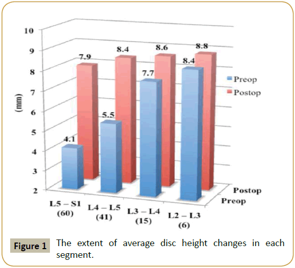
Total number of fused segments was 122 and levels of the instrumented segments were the following: L5-S1: 27; L4-S1: 18; L4-L5: 8; L3-S1: 9 and L2-S1: 6. The extent of disc height changes in each segment is shown in Figure 1.
The total length of the lumbar spine was distracted by an average 8.0 millimeters (SD: 3.7) from 194.4 (SD: 7.7) to 201.8 (SD: 8.2) mm.
MRI findings
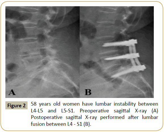
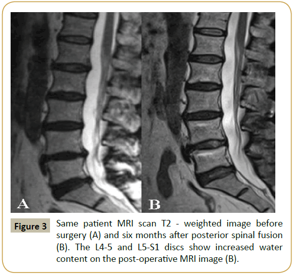
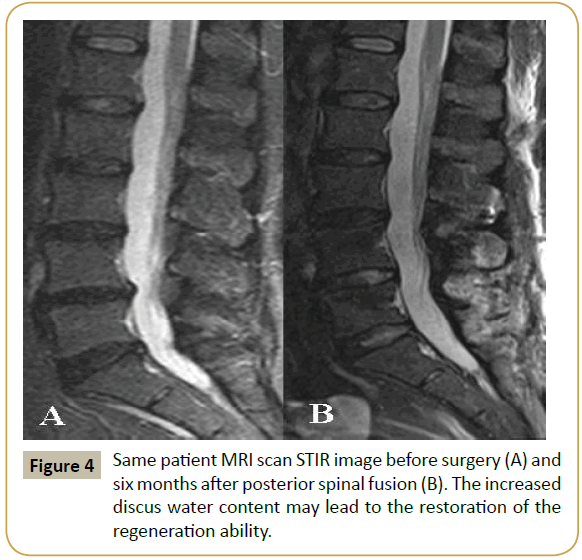
Tissue response to surgery was evident in sagittal views, mostly on T2 - weighted intensity images. Spines that were evaluated after one year of surgery demonstrated significant increase in disc hydration. After one year on MRI pictures, the water content of the distracted discs became similar to the normal discs above the lesion (Figures 2-4). At that time, neither clinical nor radiologic signs of pseudoarthrosis nor non-union of the fused spine were detected.
Functional outcome
ODI score was used preoperatively and one year after surgery for postoperative evaluation of patients’ outcomes. 61 of 68 (90%) patients had preoperative ODI. 56 of 68 (83%) patients had postoperative scores available. The average preoperative ODI score was 68.8% (SD: 6.8), which decreased to 28.4% (SD: 3.8). That represents an average of 38.2% (SD: 4.6) statistically ≤ 0 significant 0.001). Improvement (p after two-year clinical followup our patient's satisfaction did not change and did not require additional clinical or radio imaging studies.
During the follow-up period, none of our patients were reoperated on by us because of pain, implant failures, or recurrent lumbar spine problems. Also, we have no information that anyone of our patients would have been operated on in any other institutions because of problems associated with the first spinal surgery or adjacent level disease.
Discussion
Even though most spine specialists agree on the existence of lumbar spine instability, neither the definition nor the clinical picture of the disease is clearly defined [12]. Nevertheless,lumbar spine instability formed on the basis by disc degeneration is considered one of the most important factors of spinal pain and one of the most common indications for fusion surgery following ineffective conservative treatment [8].
In the third phase, so-called secondary stabilization of the disease, the clinical picture is dominated by radicular compression caused by secondary stenosis of the spinal canal developing on the basis by of repetitive rotational overload inducing increased bone turnover which eventually leads to osteophyte formation [19]. Indication for surgery is obvious with a primary aim to liberate neurological components.
In the second, unstable phase, however, the surgical indication is sometimes difficult to set up. Currently, the most common surgical procedure for segmental instability is ventral fusion accompanied with posterior transpedicular fixation [13]. During any approach of ventral fusion (PLIF, TLIF, ALIF, etc.) the intervertebral disc is always sacrificed. The question may arise, whether is not that an excessively aggressive intervention to sacrifice all discs? This question is particularly pointed in the first phase of instability called temporary dysfunction, wherever only “dehydrated” or dark discus can be found behind the complaints. Would not be a better approach to help regeneration of discus function in this stage?.
The histological and biomechanical analysis of the motion segments show that intervertebral disc damage and its progression only occurs through a "degenerative cascade", when continuous axial overloading is exerted on the discs [20]. Due to increased hydrostatic pressure on the discs water content is reduced mainly in the nucleus pulpous and the annulus fibrosus, as well, resulting in loss of the capacity for collagen and proteoglycan synthesis and regeneration of the discs, an ability that is rather slow even in healthy discs [21]. The constant disc overload progressively leads to disc degeneration, eventually causing segmental instability [22].
By pressure reduction on intervertebral discs, its regenerative capacity can be significantly improved. This could be easily obtained by intervertebral distraction [23,24]. In clinical practice, posterior lumbar distraction is not a widely accepted procedure during spinal fusion surgery. The reason for this is that posterior lumbar distraction is considered to be the key source for lumbar lordosis reduction and sagittal imbalance amplification [25]. However, this statement is based primarily on the negative results observed after using Harrington instrumentation for lumbar spine fusion, and unfortunately, is firmly established as an axiom in spine surgeons' beliefs. It is clear that posterior distraction of the lumbar spine on a straight Harrington rod will diminish lumbar lordosis and result in flat back syndrome [26].
The introduction of surgical instrumentation allowing threedimensional correction of spine deformities fundamentally altered the potential for the preservation of lumbar lordosis. With the distraction fixed on rods that are shaped corresponding to the desired postoperative lordosis, it is possible to distract adjacent vertebral bodies without diminishing the lordosis of the spine. In our series, despite posterior distraction, lumbar lordosis was not decreased; on the contrary, an average 10° increase of the lumbar spine lordosis was observed.
However, the use of monoaxial screws is primordial. A more upto- date option, the use of polyaxial screws provides the ability to alter the axis of the screw relative to its screw head. During the posterior distraction, aligned screw heads could be fixed to the pre-bended rod without displacement of adjacent screws therefore without moving away vertebral bodies above and below. In contrast, posterior distraction using monoaxial screws lead to the displacement of adjacent vertebral bodies, as a result of the fixed and constant axis of the screw and its screw head. As a consequence of intervertebral distraction, reduction of hydrostatic pressure on intervertebral discs is achieved. In this study, the posterior distraction vertebrae was carried out until intervertebral distances became comparable to normal values in healthy discs above or below.
Intervertebral disc height is partly influenced by the water content of the nucleus pulpous, which is normally about 80%. MRI provides a sensitive method to determine the water content of the intervertebral disc [27]. The decrease of the water content of nucleus pulposus indicates the progression of disc degeneration while the increase of water content reflects the result of its regenerative capacity [28]. After axial posterior distraction, postoperative MRI images revealed an increase of water content inside the disc tissue, especially inside the nucleus pulpous. In our opinion, this rise of water content may be associated with the increasing regenerative capacity of intervertebral discs.
Lumbar fusion in spinal instability cases usually leads to a significant improvement in low back pain and spinal function, as well [29].
Clinical outcome was assessed by using ODI in our study, because it is a valid condition-specific measure of spine-related disability and an objective tool to evaluate the efficacy of clinical treatment. It measures the daily functional disability induced by spine pathology. The improvement of the ODI score is a direct indicator of the impact of lumbar fusion and the decline in the patients’ symptoms [30]. An average 38% improvement of ODI score achieved in our series denotes a significant and considerable clinical improvement.
Conclusion
Posterior lumbar fusion after distraction on rods shaped corresponding to a normal degree of lordosis helps to restore the anatomic disc height and improves the quality of live in patients suffering from low back pain due to degenerative lumbar segmental instability. Adequate vertebral alignment and spinal stability is restored by surgical fusion. The MRI findings indicate that disc height restorations allow a better rehydration and probably better nutrient supply of the discs.
Our observation raises several questions.
Whether the imaging phenomenon is accompanied by any meaningful biological regeneration of intervertebral discs? This issue is currently under our research.
If any biochemical or biological signs of regeneration could be found after mechanical distraction, the following questions need to be answered: Is posterior arthrodesis necessary in all cases, or perhaps only dynamic stabilization or temporary posterior distractions in a minimally invasive way would be enough to restore the normal intervertebral disc functions?
Compliance with Ethical Standards
Conflict of Interest
The authors declare that they have no conflicts of interest that are directly or indirectly related to the research.
Ethical approval
All procedures performed in studies involving human participants were in accordance with the ethical standards of the institutional and/or national research committee and with the 1964 Helsinki declaration and its later amendments or comparable ethical standards.
References
- Manchikanti L, Singh V, Datta S, Cohen SP, Hirsch JA (2009) Comprehensive review of epidemiology, scope and impact of spinal pain. Pain Physician 12: 35-70.
- Zheng CJ, Chen J (2015) Disc degeneration implies low back pain. Theoretical Biology and Medical Modelling 12: 24.
- Iatridis JC, McLean JJ, Roughley PJ, Alini M (2006) Effects of mechanical loading on intervertebral disc metabolism in vivo. J Bone Joint Surg Am 88: 41-46.
- Battié MC, Videman T (2006) Lumbar Disc Degeneration: Epidemiology and Genetics. J Bone Joint Surg Am88: 3-9.
- Kirkaldy-Willis WH, Wedge JH, Yong-Hing K, Tchang S, de Korompay V, et al. (1982) Instability of the lumbar spine. Clinical Orthopaedics and Related Research 165: 110-123.
- Haughton V (2011) The “dehydrated” lumbar intervertebral disk on MR, its anatomy, biochemistry and biomechanics. Neuroradiology 53: S191-S194.
- Borenstein DG, O’Mara JW, Boden SD, Lauerman WC, Jacobson A, et al. (2001) The value of magnetic resonance imaging of the lumbar spine to predict low back pain in asymptomatic subjects: A 7-year follow-up study. J Bone Joint Surg Am 83: 1306-1311.
- Leone A, Guglielmi G, Cassar-Pullicino VN, Bonomo L (2007) Lumbar intervertebral instability. A Review. Radiology 245: 62-77.
- Deyo RA (1993) Practice variations, treatment fads, rising disability: Do we need a new clinical research paradigm? Spine 18: 2153-2162.
- Modic MT, Ross JS (2007) Lumbar degenerative disk disease. Radiology 245: 43-61.
- Kettler A, Rohlmann F, Ring C, Mack C, Wilke HJ (2011) Do early stages of lumbar intervertebral disc degeneration really cause instability? Evaluation of an in vitro database. Eur Spine J 20: 578-584.
- Colaiacomo MC, Tortora A, Di Biasi C, Polettini E, Casciani E, et al. (2009) Intervertebral instability. Clin Ther 160: 75-82.
- Lykissas MG, Aichmair A (2013) Current concepts on spinal arthrodesis in degenerative disorders of the lumbar spine. World J Clin Cases 1: 4-12.
- Yoshihara H, Yoneoka D (2015) National trends in the surgical treatment for lumbar degenerative disc disease. Spine J 15: 265-271.
- Tibiletti M, Kregar Velikonja N, Urban JPG, Fairbank JCT (2014) Disc cell therapies: critical issues. Eur Spine J 23: 375-384.
- Hee HT, Chuah YJ, Tan BH, Setiobudi T, Wong HK (2011) Vascularisation and morphological changes of the endplates after axial compression and distraction of the intervertebral disc” Spine 36: 505-511.
- Kroeber M, Unglaub F, Guehring T, Nerlich A, Hadi T, et al. (2005) Effects of Controlled Dynamic Disc Distraction on Degenerated Intervertebral Discs. An in Vivo Study on the Rabbit Lumbar Spine Model. Spine 30: 181-187.
- Fairbank JCT, Pynsent PB (2000) The oswestry disability index. Spine 25: 2940-2953.
- Farfan HF (1980) The pathological anatomy of degenerative spondylolisthesis. A cadaver study. Spine 5: 412-418.
- Stokes IAF, Latridis JC (2004) Mechanical Conditions That Accelerate Intervertebral Disc Degeneration: Overload Versus Immobilization. Spine 29: 2724-2732.
- Lotz JC, Colliou OK, Chin JR, Duncan NA, Liebenberg E (1998) Compression-induced degeneration of the intervertebral disc: an in vivo mouse model and finite-element study. Spine 23: 2493-2506.
- Adams MA, Roughley PJ (2006) What is Intervertebral Disc Degeneration, and What Causes It? Spine 31: 2151-2161.
- Guehring T, Unglaub F, Lorenz H, Omlor G, Wilke HJ, et al. (2006) Intradiscal pressure measurements in normal discs, compressed discs and compressed discs treated with axial posterior disc distraction: an experimental study on the rabbit lumbar spine model. Eur Spine J 15: 597-604.
- Guehring T, Omlor GW, Lorenz H, Engelleiter, K, Richter W, et al. (2006) Disc distraction shows evidence of regenerative potential in degenerated intervertebral discs as evaluated by protein expression, magnetic resonance imaging, and messenger ribonucleic acid expression analysis. Spine 31: 1658-1665.
- Potter BK, Lenke LG, Kuklo TR (2004) Prevention and Management of Iatrogenic Flatback Deformity. J Bone Joint Surg Am 86: 1793-1808.
- Lu DC, Chou D (2007) Flatback syndrome. Neurosurg Clin N Am 18: 289-295.
- Marinelli NL, Haughton VM, Munoz A, Anderson PA (2009) T2 relaxation times of intervertebral dis tissue correlated with water content and proteoglycan content. Spine 34: 520-524.
- Chiu EJ, Newitt DC, Segal MR, Hu SS, Lotz JC, et al. (2001) Magnetic resonance imaging measurement of relaxation and water dissucion in the human lumbar intervertebral disc under compression in vitro. Spine 26: 437-44.
- Phillips FM, Slosar PJ, Youssef JA, Andersson G, Papatheofanis F (2013) Lumbar spine fusion for chronic low back pain due to degenerative disc disease: a systematic review. Spine 38: 409-422.
- Carreon LY, Glassman SD, Howard J (2008) Fusion and non-surgical treatment for symptomatic lumbar degenerative disease: a systematic review of Oswestry Disability Index and MOS Short Form-36 outcomes. Spine J 8: 747-755.
Open Access Journals
- Aquaculture & Veterinary Science
- Chemistry & Chemical Sciences
- Clinical Sciences
- Engineering
- General Science
- Genetics & Molecular Biology
- Health Care & Nursing
- Immunology & Microbiology
- Materials Science
- Mathematics & Physics
- Medical Sciences
- Neurology & Psychiatry
- Oncology & Cancer Science
- Pharmaceutical Sciences
